Table of Contents >> Show >> Hide
- What Are Lumbar Lymph Nodes?
- Lumbar Lymph Nodes Anatomy: Where They Are Located
- How to Understand a Lumbar Lymph Nodes Diagram
- What Do the Lumbar Lymph Nodes Drain?
- Function of the Lumbar Lymph Nodes
- Why Lumbar Lymph Nodes Matter Clinically
- Lumbar Lymph Nodes and Cancer Spread
- How Doctors Evaluate Lumbar Lymph Nodes
- Key Takeaways About Lumbar Lymph Nodes
- What People Often Experience When This Anatomy Becomes Relevant
- Conclusion
Deep in the back of the abdomen, tucked beside the body’s biggest blood vessels, the lumbar lymph nodes do some of their best work without asking for applause. They are part of the retroperitoneal lymphatic system, which is a fancy way of saying they live behind the lining of the abdominal cavity and help manage fluid drainage, immune surveillance, and the spread of disease. They are not celebrity lymph nodes like the ones in the neck or armpit that everyone can feel when they swell. These are the behind-the-scenes professionals.
If you have ever seen the terms lumbar lymph nodes, para-aortic lymph nodes, periaortic nodes, or retroperitoneal lymph nodes, welcome to the anatomical naming maze. Some sources use these labels a little differently. In everyday medical discussion, lumbar lymph nodes usually refer to the deep lymph-node chains around the abdominal aorta and inferior vena cava. In practice, the most talked-about group is the para-aortic group, because it matters in abdominal, pelvic, gynecologic, renal, and testicular disease.
This guide breaks down where the lumbar lymph nodes live, what a typical diagram is showing, what they drain, why they matter in real life, and what people may experience when these nodes become enlarged or clinically important.
What Are Lumbar Lymph Nodes?
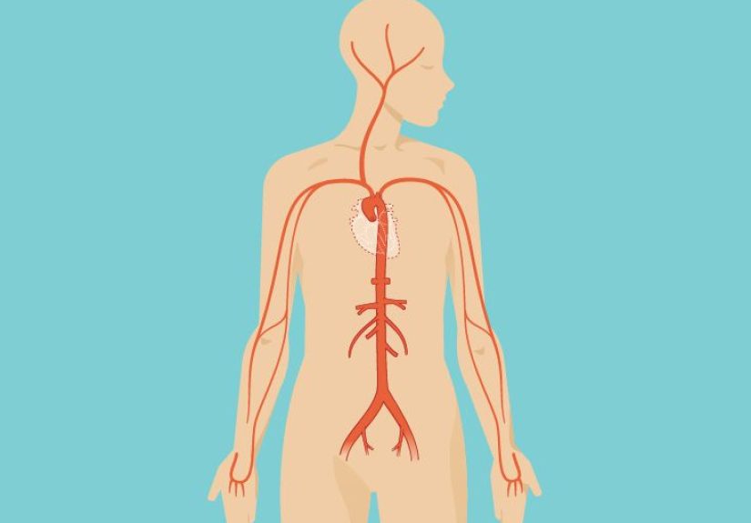
Lumbar lymph nodes are a cluster of deep lymph nodes in the retroperitoneum, the space behind the peritoneum. They lie along the abdominal aorta and the inferior vena cava, generally extending from the upper abdomen down toward the aortic bifurcation and common iliac region. Because they sit alongside the great vessels, they are strategically positioned to receive lymph from major abdominal and pelvic structures.
Think of them as a quality-control checkpoint for lymph coming from organs and tissues that do not get much public attention until something goes wrong. Their job is to filter lymph, trap harmful particles, and coordinate immune responses. They also serve as an anatomical roadmap for how certain cancers spread. That last role is why doctors care so much about them on CT scans, MRIs, PET scans, and during surgeries such as retroperitoneal lymph node dissection.
Lumbar Lymph Nodes Anatomy: Where They Are Located
From an anatomy standpoint, the lumbar lymph nodes are grouped around the body’s large abdominal vessels. A broader description divides them into left lumbar, intermediate lumbar, and right lumbar chains. A more detailed description names them by their relationship to the aorta or inferior vena cava.
Groups Around the Aorta
- Pre-aortic nodes: Located in front of the aorta. These are associated with lymph drainage from many abdominal digestive organs.
- Para-aortic nodes: Located beside the aorta. These are commonly called lumbar aortic nodes or lateral aortic nodes.
- Retro-aortic nodes: Located behind the aorta.
Groups Around the Inferior Vena Cava
- Pre-caval nodes: In front of the inferior vena cava.
- Para-caval nodes: Beside the inferior vena cava.
- Retro-caval nodes: Behind the inferior vena cava.
Interaortocaval or Intermediate Nodes
Some lymph nodes sit in the narrow zone between the aorta and the inferior vena cava. These are often called interaortocaval or intermediate lumbar nodes. Clinically, they are especially important in the spread pattern of testicular cancer, particularly on the right side.
These nodes are closely related to nearby landmarks such as the kidneys, renal vessels, adrenal glands, psoas major muscles, diaphragmatic crura, ureters, and common iliac vessels. In other words, they live in a crowded neighborhood with high-stakes neighbors.
How to Understand a Lumbar Lymph Nodes Diagram
If you are looking at a diagram of the lumbar lymph nodes, do not let the arrows and labels bully you. Most diagrams are trying to show the same basic idea: a vertical lymphatic highway wrapped around the abdominal aorta and inferior vena cava.
A Good Diagram Usually Labels These Structures
- The abdominal aorta in the center-left of the retroperitoneum
- The inferior vena cava to the right of the aorta
- The kidneys and adrenal glands higher up
- The renal vessels crossing near the upper para-aortic region
- The common iliac vessels lower down near the aortic bifurcation
- The psoas major muscles running vertically on each side
- The cisterna chyli or upper lumbar trunks, where filtered lymph is funneled upward toward the thoracic duct
What the Diagram Is Really Telling You
A lumbar lymph node diagram is not just a geography lesson. It is showing how lymph travels. Nodes beside the aorta collect drainage from organs like the kidneys, adrenal glands, ovaries, testes, uterine tubes, and parts of the uterus. Nodes in front of the aorta handle drainage from digestive organs. Nodes near the vena cava and between the great vessels connect these routes and help move lymph upward.
So when a radiology report mentions para-aortic adenopathy or retroperitoneal lymphadenopathy, it is pointing to this deep-node system. That can matter for infection, inflammation, lymphoma, or metastatic cancer.
What Do the Lumbar Lymph Nodes Drain?
This is where anatomy gets practical. The lumbar lymph nodes are not random beans in the abdomen. They receive lymph from very specific territories.
Major Drainage Areas
- Kidneys and suprarenal glands: These structures commonly drain to para-aortic nodes.
- Posterior abdominal wall: Deep lymphatics from the posterior wall can reach lateral aortic and retro-aortic nodes.
- Common iliac chain: Lymph from the pelvis and lower limbs can move upward through iliac nodes before reaching the lumbar chain.
- Testes: Testicular lymphatics follow the gonadal vessels to retroperitoneal para-aortic nodes rather than to superficial groin nodes.
- Ovaries: Ovarian lymph typically travels with the ovarian vessels to para-aortic nodes, often near the level of the renal vessels.
- Fallopian tubes: Drainage may reach both para-aortic and pelvic lymph nodes.
- Uterine fundus: The top of the uterus can drain to para-aortic nodes, while lower portions usually drain more toward pelvic nodes.
- Digestive organs: Pre-aortic nodes receive drainage from the GI tract, spleen, pancreas, gallbladder, and liver.
This pattern explains one of the most important clinical pearls in anatomy: the testes do not primarily drain to the scrotal or inguinal nodes. They drain upward to retroperitoneal para-aortic nodes because of their embryologic origin near the kidneys. That single fact has saved many anatomy students from exam heartbreak and helps doctors predict where disease may spread.
Function of the Lumbar Lymph Nodes
The lumbar lymph nodes function can be grouped into three big jobs: filtration, immune defense, and transport.
1. Filtering Lymph
Lymph nodes filter lymphatic fluid before it is returned to the bloodstream. As lymph passes through the node, immune cells inspect it for bacteria, viruses, damaged cells, and potentially malignant cells. It is the medical version of airport security, except the staff is made of lymphocytes and macrophages.
2. Supporting Immune Surveillance
These nodes help the immune system recognize threats and mount responses. If infection or inflammation is present in tissues that drain to the lumbar chain, these nodes may enlarge as immune activity increases.
3. Moving Lymph Upward
After filtration, lymph leaves through efferent vessels that contribute to the right and left lumbar trunks. These trunks usually connect with the cisterna chyli, which then continues as the thoracic duct. This pathway returns filtered lymph back to the venous circulation.
In plain English, the lumbar nodes are both a checkpoint and a transfer station. They inspect the cargo, then help send it on its way.
Why Lumbar Lymph Nodes Matter Clinically
Because these nodes are deep, they cannot usually be felt from the outside. That means problems involving them are often detected with imaging rather than with a routine physical exam. When they enlarge, the causes range from routine to serious.
Common Clinical Scenarios
- Infection: Lymph nodes may swell in response to infection, though deep retroperitoneal node enlargement is not something most people notice directly.
- Inflammation: Autoimmune and inflammatory processes can involve abdominal lymph nodes.
- Lymphoma: Cancers of the lymphatic system can enlarge retroperitoneal nodes.
- Metastatic cancer: Testicular, ovarian, uterine, cervical, renal, gastrointestinal, pancreatic, and other cancers may spread to para-aortic or retroperitoneal nodes.
- Cancer staging and treatment planning: Their involvement can change staging, treatment decisions, radiation fields, and surgical plans.
Symptoms When Lumbar or Retroperitoneal Nodes Enlarge
Enlarged lumbar lymph nodes may cause deep abdominal discomfort, bloating, or lower back pain. Some people have no obvious symptoms and only learn about enlarged retroperitoneal nodes after a CT scan done for another reason. Others may develop symptoms related to the underlying disease rather than the nodes themselves, such as fever, night sweats, weight loss, pelvic symptoms, urinary issues, or cancer-specific warning signs.
Since these nodes sit near ureters, vessels, nerves, and major organs, large masses in this region can sometimes create pressure effects. That is one reason retroperitoneal disease gets taken seriously even when the first symptom seems vague.
Lumbar Lymph Nodes and Cancer Spread
One reason this anatomy gets so much attention is that cancer does not spread randomly. It often follows lymphatic routes.
Testicular Cancer
This is the classic example. The testes develop near the kidneys during fetal life, so their lymphatic drainage follows the gonadal vessels back to retroperitoneal para-aortic nodes. That is why doctors may examine the retroperitoneum carefully in patients with testicular cancer, and why retroperitoneal lymph node dissection can be part of management.
Gynecologic Cancers
Ovarian, uterine, tubal, and cervical cancers may involve para-aortic nodes. In some gynecologic cancers, para-aortic nodal status helps determine stage and treatment. These nodes may also be included in radiation planning or surgical staging, depending on the disease.
Abdominal and Pelvic Cancers
Kidney, pancreatic, colorectal, gastric, and other abdominal cancers may spread through regional lymphatic chains that connect with retroperitoneal nodes. For radiologists and oncologists, lumbar-node anatomy is not academic trivia. It is the map.
How Doctors Evaluate Lumbar Lymph Nodes
Because the nodes are deep, evaluation usually relies on imaging and context rather than touch alone.
Common Tools
- CT scan: Frequently used to identify enlarged retroperitoneal nodes and define their relationship to vessels and organs.
- MRI: Helpful in selected cases, especially when soft-tissue detail matters.
- PET scan: Can help evaluate metabolic activity when cancer is suspected.
- Biopsy or dissection: Sometimes needed to confirm diagnosis or stage disease.
Radiology reports may use terms like lymphadenopathy, para-aortic nodes, periaortic nodes, interaortocaval nodes, or retroperitoneal adenopathy. These labels help pinpoint exactly which node groups are involved.
Key Takeaways About Lumbar Lymph Nodes
The lumbar lymph nodes are deep retroperitoneal nodes arranged around the abdominal aorta and inferior vena cava. They collect lymph from major abdominal and pelvic organs, filter it, support immune defense, and pass it upward through the lumbar trunks toward the cisterna chyli and thoracic duct. Their anatomy matters because disease often follows the lymphatic map, especially in cancers involving the testes, ovaries, uterus, kidneys, and other abdominal organs.
In short, these nodes are small, quiet, and absolutely not messing around.
What People Often Experience When This Anatomy Becomes Relevant
For most people, the lumbar lymph nodes remain invisible and anonymous for life. Nobody wakes up thinking, “I wonder how my para-aortic chain is doing today?” Yet these nodes suddenly become very relevant in a few common clinical situations, and the experience can be confusing because the symptoms are often indirect.
One common experience is the accidental discovery. A person has abdominal pain, back pain, kidney-stone symptoms, unexplained bloating, or a follow-up scan after surgery, and the radiology report mentions enlarged retroperitoneal or para-aortic lymph nodes. That moment can feel alarming because the phrase sounds dramatic and deeply anatomical, which it is. But the finding itself is not a diagnosis. It is a clue. The next step is figuring out whether the nodes are reacting to infection, inflammation, lymphoma, or metastatic disease, or whether they simply need interval follow-up imaging.
Another experience is deep, vague discomfort. Unlike a swollen lymph node in the neck, lumbar lymph nodes cannot be poked with a fingertip in front of a mirror. When these deep nodes enlarge, the sensation is often described less as “I can feel a lump” and more as “something feels off in my lower back or belly.” Some people report pressure, fullness, or pain that seems hard to pinpoint. Because the retroperitoneum houses major vessels, ureters, nerves, and connective tissue, symptoms may come from local pressure rather than from the node itself.
In cancer care, the experience is often about staging and planning. A patient with testicular, ovarian, uterine, cervical, or kidney cancer may hear that doctors are checking para-aortic nodes to see whether disease has spread. That information can influence whether treatment includes surgery, chemotherapy, radiation, surveillance, or some combination of the above. In this setting, lumbar-node anatomy becomes more than a textbook topic. It becomes part of decision-making, prognosis, and the emotional language of a diagnosis.
For some patients, the experience centers on surgery or treatment discussions. A surgeon may explain a retroperitoneal lymph node dissection, while a radiation oncologist may discuss targeting para-aortic nodes in the abdomen. These conversations can sound intimidating, mostly because the area is deep and surrounded by important structures. Understanding the anatomy helps patients ask better questions: Which node groups are involved? Are the nodes enlarged on imaging, positive on biopsy, or being treated because they are part of a known drainage pathway? Is the goal diagnosis, staging, treatment, or all three?
There is also the experience of uncertainty. Enlarged lymph nodes are not automatically cancer, and painful nodes are not automatically harmless. Some inflammatory or infectious causes improve. Some findings remain stable and are just monitored. Others need more urgent workup. That gray zone is frustrating, but it is common. The anatomy matters because it helps clinicians narrow the possibilities. Nodes near the aorta and vena cava are not random; they reflect drainage from specific organs and tissues, which gives doctors a logical place to start.
The most helpful takeaway for patients and families is this: if lumbar lymph nodes show up in a report, ask what they likely drain, whether the enlargement is mild or significant, what other findings are present, and what the next step is. The anatomy may be deep, but the explanation should not be.
Conclusion
The lumbar lymph nodes may be buried in the retroperitoneum, but they are central to how the body filters lymph, coordinates immune defense, and reveals patterns of disease spread. Understanding their location around the aorta and inferior vena cava, their drainage territories, and their role in imaging and cancer staging makes the whole abdomen make a lot more sense. Whether you are reading a scan report, studying anatomy, or trying to decode a confusing medical term, knowing the lumbar lymph nodes is like finally getting the legend on the map.