Table of Contents >> Show >> Hide
- What Are Supraclavicular Lymph Nodes?
- Where Are the Supraclavicular Lymph Nodes Located?
- Simple Diagram of Supraclavicular Lymph Node Drainage
- What Do Supraclavicular Lymph Nodes Do?
- Why Are These Nodes So Clinically Important?
- How Doctors Examine Supraclavicular Lymph Nodes
- Supraclavicular Lymph Nodes and Virchow’s Node
- Symptoms People May Notice
- When to Seek Medical Attention
- Real-World Experiences Related to Supraclavicular Lymph Nodes
- Conclusion
Supraclavicular lymph nodes are tiny structures with a very un-tiny reputation. They sit just above the collarbone, quietly filtering lymph, helping the immune system spot trouble, and generally doing their best “security checkpoint” impression. Most days, they work without applause. But when one becomes enlarged, doctors pay attention fast, because this region can offer important clues about infection, inflammation, or sometimes a more serious underlying condition.
If the name sounds like it was invented by someone who loved crossword puzzles, here is the simple version: supra means above, and clavicular refers to the clavicle, or collarbone. So these are the lymph nodes above the collarbone. Easy. Their location may be small on the body map, but their clinical importance is anything but small. In anatomy, they are part of the lower neck lymphatic network. In medicine, they are famous because the right and left sides can hint at different drainage pathways and different disease patterns.
This guide breaks down the anatomy, function, drainage, and real-world meaning of supraclavicular lymph nodes in plain American English. You will also get a simple diagram-style explanation, because nobody should need an advanced anatomy lab just to understand the base of the neck.
What Are Supraclavicular Lymph Nodes?
Supraclavicular lymph nodes are part of the lymphatic system, the body’s cleanup crew and infection patrol rolled into one. Like other lymph nodes, they are small, bean-shaped filters made of lymphoid tissue. Lymph, a clear fluid rich in immune cells, travels through lymphatic vessels and passes through nodes before returning to the bloodstream. Along the way, the nodes trap bacteria, viruses, abnormal cells, and cellular debris.
These nodes belong to the cervical lymph node region, specifically near the lower neck and the supraclavicular fossa, which is the hollow just above the clavicle. They are closely related to nearby structures such as the sternocleidomastoid muscle, major veins at the base of the neck, and the terminal pathways where lymph rejoins the circulation.
That is why supraclavicular nodes matter so much. They are not random bumps in random places. They sit near one of the body’s major lymphatic crossroads.
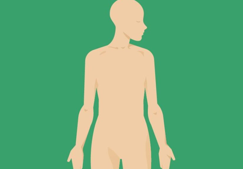
Where Are the Supraclavicular Lymph Nodes Located?
Anatomically, the supraclavicular lymph nodes lie just superior to the clavicle at the base of the neck. You can think of them as living in the shallow valley where the neck meets the shoulder. They are often grouped with the lower deep cervical lymphatic chain, and they are close to the venous angle where major veins and lymphatic ducts meet.
Their position matters because the lymphatic system is directional. Lymph does not wander around like a tourist without a map. It follows organized routes. The supraclavicular area acts like a final checkpoint before lymph empties into the bloodstream.
Right Supraclavicular Lymph Nodes
The right supraclavicular region is associated with drainage from the right side of the head and neck, the right upper chest, and the right upper extremity through the right lymphatic duct. Because of this anatomy, an enlarged right supraclavicular node can sometimes be linked to conditions involving the mediastinum, lungs, or esophagus. That does not mean every right-sided lump is a cancer alarm bell, but it does mean the finding should not be shrugged off like a sock under the couch.
Left Supraclavicular Lymph Nodes
The left supraclavicular region is especially famous because it sits near the end of the thoracic duct, the body’s largest lymphatic vessel. The thoracic duct drains lymph from a much broader territory, including the left side of the head and neck, the left chest, the abdomen, the pelvis, and both legs. Because of this, the left supraclavicular node may become enlarged when disease from the chest or even below the diaphragm travels through the lymphatic system.
The best-known left-sided node is Virchow’s node. It is classically described as a left supraclavicular node near the junction of the thoracic duct and the left venous angle. When enlarged due to metastatic disease, it has historically been associated with cancers of the stomach and other abdominal or pelvic organs, though it can also be involved in thoracic malignancies and lymphoma.
Simple Diagram of Supraclavicular Lymph Node Drainage
Here is a plain-language diagram to make the anatomy easier to picture:
Head and neck tissues → cervical lymphatic chains → supraclavicular region → major lymphatic ducts → venous circulation
Right side drainage:
Right head/neck + right chest + right arm → right lymphatic duct → right venous angle near the base of the neck
Left side drainage:
Left head/neck + left chest + abdomen + pelvis + both legs → thoracic duct → left venous angle near the base of the neck
Clinical pearl:
Enlarged left supraclavicular node = sometimes linked to disease from the chest or abdomen
Enlarged right supraclavicular node = sometimes linked to disease in the right thorax, mediastinum, or esophagus
This is a conceptual diagram, not a surgical map. But it captures the big idea: supraclavicular nodes sit at a drainage intersection, which is why they can reflect disease from more distant body regions than many people expect.
What Do Supraclavicular Lymph Nodes Do?
The function of supraclavicular lymph nodes is the same as the function of lymph nodes elsewhere, but their location gives them extra diagnostic significance.
1. They Filter Lymph
Lymph nodes act as biological filters. As lymph passes through, immune cells inside the node inspect it for pathogens, damaged tissue, and abnormal cells. That means supraclavicular nodes help clean lymph before it rejoins the bloodstream.
2. They Support Immune Surveillance
These nodes contain lymphocytes and other immune cells that identify and respond to infections. If your body is fighting a nearby or systemic infection, the nodes may enlarge because immune activity ramps up. In plain English, they get busy and a little grumpy-looking.
3. They Help Signal Disease Patterns
This is where supraclavicular nodes become medically famous. Because of their drainage relationships, enlargement here can reflect local infections, inflammatory disease, lymphoma, or metastatic spread from distant organs. A node in this location is not merely a swollen gland; it can be an anatomical clue.
Why Are These Nodes So Clinically Important?
Doctors tend to be more cautious about supraclavicular lymphadenopathy than enlargement in many other areas. That is because supraclavicular nodes have a higher association with malignancy, especially when they are firm, fixed, painless, or persistent. Age over 40, systemic symptoms such as night sweats or unexplained weight loss, and a node that stays enlarged for more than a couple of weeks can all raise concern.
Still, context matters. Painful, suddenly enlarged lymph nodes are more often related to infection or inflammation. Slow, painless enlargement deserves closer evaluation. In short, a tender node after an infection is one story; a hard, persistent node with no obvious cause is another story entirely.
Common Benign Causes
Not every enlarged supraclavicular node means cancer. Benign causes can include upper respiratory infections, viral illness, bacterial infections, inflammatory conditions, and occasionally reactive changes after the immune system has been stimulated. That said, because of the location, clinicians usually do not stop at “probably nothing” without a good reason.
Common Serious Causes
More serious causes include lymphoma, metastatic cancer, and less commonly other systemic diseases. The classic teaching is that the left supraclavicular node may point toward thoracic or abdominal malignancy, while the right side may be associated more often with disease in the chest, mediastinum, or esophagus. These are patterns, not iron laws, but they are patterns physicians take seriously.
How Doctors Examine Supraclavicular Lymph Nodes
On physical exam, the supraclavicular area is usually checked with the patient sitting upright. The examiner gently palpates the hollows above the clavicles, comparing both sides. Sometimes a patient may be asked to relax the shoulders or take a breath in a certain way to make subtle nodes easier to feel.
Doctors pay attention to several features:
Size: Bigger nodes are generally more concerning than tiny, barely perceptible ones.
Tenderness: Tender nodes often suggest infection or inflammation.
Consistency: Hard nodes can be more worrisome than soft, mobile nodes.
Mobility: Fixed nodes raise more concern than freely movable ones.
Duration: A node that persists or grows over time deserves evaluation.
Associated symptoms: Fever, weight loss, cough, trouble swallowing, fatigue, or night sweats can guide the workup.
If the finding remains suspicious, the next steps may include ultrasound, CT imaging, laboratory testing, or tissue sampling with fine-needle aspiration or biopsy. The goal is not to panic; the goal is to identify the cause accurately.
Supraclavicular Lymph Nodes and Virchow’s Node
No discussion of supraclavicular lymph nodes is complete without Virchow’s node, the celebrity guest star of neck anatomy. Virchow’s node is the classic left supraclavicular node associated with metastatic spread through the thoracic duct. Because the thoracic duct collects lymph from much of the body below the diaphragm, enlargement in this area can sometimes reflect disease originating far away from the neck itself.
Historically, clinicians have linked Virchow’s node with gastric cancer, but the association is broader than that. Other abdominal, pelvic, thoracic, and lymphoid malignancies may also present this way. In other words, Virchow’s node is less a one-hit wonder and more a reminder that anatomy and pathology love to collaborate.
The practical takeaway is simple: a palpable left supraclavicular node, especially if firm and persistent, is not something to monitor casually without medical input.
Symptoms People May Notice
Some people never notice these nodes at all unless a clinician points them out. Others feel a lump above the collarbone while applying lotion, shaving, getting dressed, or absentmindedly poking at their neck during a stressful work call. The lump may feel tender and sore, or it may feel firm and painless.
Other symptoms depend on the cause. If infection is the trigger, a person may also have fever, sore throat, recent illness, or localized pain. If a systemic illness is involved, symptoms may include fatigue, night sweats, appetite loss, unexplained weight loss, cough, gastrointestinal complaints, or a general sense that something is off.
The location of the node does not diagnose the cause by itself, but it does shape the questions doctors ask next.
When to Seek Medical Attention
A healthcare evaluation is a smart move if you notice a supraclavicular lump that is new, getting bigger, or not clearly tied to a recent infection. Evaluation is especially important if the node is hard, fixed, painless, or accompanied by fever, night sweats, unintended weight loss, persistent cough, chest symptoms, or abdominal symptoms.
This is one of those body findings where “I’ll just keep an eye on it for the next six months” is not the winning strategy. Most causes are still diagnosable and manageable, but the location earns it an earlier conversation with a clinician.
Real-World Experiences Related to Supraclavicular Lymph Nodes
People often encounter supraclavicular lymph nodes in surprisingly ordinary moments. One person feels a tender bump above the collarbone while getting ready for work after a week of sore throat and congestion. Another notices a painless fullness while looking in the mirror and cannot decide whether it is muscle, posture, or something new. Someone else hears the term for the first time during a breast exam, a cancer workup, or a CT scan report, and suddenly anatomy becomes very personal.
A common experience is anxiety fueled by location. Most people have heard that neck lumps can happen with infections, but the phrase “supraclavicular node” tends to make the room feel smaller. That reaction is understandable. Clinicians often respond by putting the finding into context: when did it appear, does it hurt, is it soft or hard, have there been recent infections, are there symptoms like fever or weight loss, and are there other enlarged nodes elsewhere? Those questions are not random. They reflect the way supraclavicular lymph nodes sit at the intersection of anatomy and diagnosis.
Another common experience is the difference between tender and painless swelling. A tender node after an upper respiratory infection often feels like the body is sounding an alarm while the immune system does its job. People describe it as sore when they turn the neck, wear a backpack strap, or press on the area. In contrast, a painless node can be more unsettling because it does not announce itself with inflammation. It is just there, quietly refusing to explain itself.
Patients also frequently describe the workup as a mix of relief and frustration. Relief, because the finding is being taken seriously. Frustration, because the path from lump to answer may involve several steps: physical exam, lab work, imaging, recheck visits, and sometimes biopsy. From the patient side, that can feel like a long journey for something the size of a bean. From the medical side, it is careful pattern recognition. The node is small, but the list of possible causes is large.
There are also practical day-to-day experiences. People worry about touching the area too much. They compare one side to the other in the mirror. They search online and immediately meet dramatic worst-case scenarios, which is rarely calming. Good clinical care helps here by separating possibilities into categories: reactive, infectious, inflammatory, lymphoproliferative, or metastatic. That framework does not remove uncertainty instantly, but it turns panic into process.
For clinicians, supraclavicular nodes are memorable because they can redirect an entire exam. A small node above the clavicle may prompt a more detailed review of respiratory symptoms, gastrointestinal history, breast findings, or systemic symptoms. It can change what imaging is ordered and how urgently follow-up happens. In that sense, the experience is not only emotional for patients; it is diagnostically meaningful for providers.
The most grounded way to think about these nodes is this: they are not automatically dangerous, but they are rarely ignored. A painful node after infection may settle down with time. A persistent or unexplained node deserves investigation. That balance between caution and calm is the real-world experience many people remember most. Tiny structure, big conversation.
Conclusion
Supraclavicular lymph nodes are small lymphatic filters located just above the collarbone, but their importance far exceeds their size. Anatomically, they sit at the base of the neck near major lymphatic drainage pathways. Functionally, they help filter lymph and support immune surveillance. Clinically, they matter because the right and left sides reflect different drainage territories, with the left side, especially Virchow’s node, holding special significance due to its relationship with the thoracic duct.
The big lesson is not to fear every lump, but not to dismiss this location either. Supraclavicular nodes can enlarge for benign reasons such as infection, yet persistent, hard, fixed, or painless enlargement deserves prompt medical evaluation. When it comes to anatomy, these nodes are modest. When it comes to diagnostic value, they punch well above their weight.