Table of Contents >> Show >> Hide
- What Is a Chest Wall Tumor?
- Types of Chest Wall Tumors
- Common Symptoms of Chest Wall Tumors
- What Causes Chest Wall Tumors?
- How Chest Wall Tumors Are Diagnosed
- Treatment Options for Chest Wall Tumors
- Recovery and Follow-Up Care
- When to See a Doctor
- Living With a Chest Wall Tumor Diagnosis
- Practical Experiences and Real-Life Lessons About Chest Wall Tumors
- Conclusion
Chest wall tumors sound like something that belongs in a medical drama right before the dramatic music kicks in. In real life, though, they are a complex but understandable group of growths that can develop in the bones, cartilage, muscles, soft tissues, nerves, or blood vessels that make up the chest wall. The chest wall is the sturdy “body armor” around your heart, lungs, major blood vessels, and upper abdominal organs. When a tumor forms there, doctors take it seriously because the area is both protective and mechanically important for breathing.
The good news is that not every chest wall tumor is cancer. Some are benign, slow-growing, and discovered almost by accident during imaging for something else. Others are malignant and require a coordinated treatment plan involving thoracic surgeons, oncologists, radiologists, pathologists, and sometimes plastic or reconstructive surgeons. In other words, it is not a one-doctor, one-hammer situation. It is more like assembling a highly trained medical Avengers team, minus the capes.
This guide explains the major types of chest wall tumors, common symptoms, possible causes and risk factors, how they are diagnosed, and what treatment may involve. It is written for readers who want clear, practical information without needing a medical dictionary in one hand and an espresso in the other.
What Is a Chest Wall Tumor?
A chest wall tumor is an abnormal growth that develops in or spreads to the structures of the chest wall. These structures include the ribs, sternum, cartilage, muscles, connective tissue, skin, blood vessels, and nerves. Tumors may be primary, meaning they begin in the chest wall itself, or secondary, meaning they spread to the chest wall from another cancer, such as breast cancer, lung cancer, lymphoma, kidney cancer, or other metastatic disease.
Primary chest wall tumors are relatively rare. Because of that, they are often evaluated at centers with experience in thoracic surgery and sarcoma care. Secondary chest wall involvement is more common than primary disease, especially when cancers from nearby organs grow directly into the chest wall or when cancer spreads through the bloodstream or lymphatic system.
Tumors can also be classified as benign or malignant. Benign tumors do not invade nearby tissues in the same aggressive way cancer does and usually do not spread to distant parts of the body. Malignant tumors, however, can grow into surrounding tissues, recur after treatment, and spread elsewhere if not controlled.
Types of Chest Wall Tumors
Chest wall tumors are not one single disease. They are a family reunion of very different conditions, and some relatives are much more troublesome than others. The exact type matters because treatment depends heavily on the tumor’s origin, grade, size, location, and whether it has spread.
Benign Chest Wall Tumors
Benign tumors may arise from bone, cartilage, fibrous tissue, fat, nerves, or blood vessels. Some remain stable for years and cause no symptoms. Others can grow large enough to press on nearby structures or cause pain.
Osteochondroma is one of the most common benign bone tumors. It is made of bone and cartilage and may appear as a bony bump near a rib or other skeletal structure. Many osteochondromas are monitored unless they cause pain, limit movement, irritate surrounding tissue, or show concerning changes.
Chondroma is a benign cartilage tumor. In the chest wall, cartilage tumors require careful evaluation because some malignant cartilage tumors can look deceptively calm at first. Doctors may recommend imaging, biopsy, or surgical removal depending on the lesion’s appearance and symptoms.
Fibrous dysplasia occurs when normal bone is replaced by fibrous tissue. It may cause bone expansion, deformity, pain, or fracture risk, though some cases are found incidentally. When it affects the ribs, it can appear as a chest wall mass or abnormality on imaging.
Lipoma is a benign fatty tumor. A lipoma near the chest wall often feels soft and movable, although deeper lipomas may require imaging to confirm what they are. Most are harmless, but rapid growth, pain, or unusual firmness deserves medical attention.
Neurogenic tumors arise from nerves. Many are benign, but because nerves run through tight spaces in the chest, even benign growths may cause pain, tingling, or pressure symptoms.
Malignant Chest Wall Tumors
Malignant chest wall tumors are cancers that may begin in bone, cartilage, or soft tissue. They are often grouped under sarcomas when they arise from connective tissues such as bone, cartilage, muscle, fat, or fibrous tissue.
Chondrosarcoma is one of the most common primary malignant chest wall tumors in adults. It begins in cartilage-producing cells and may involve the ribs or sternum. Chondrosarcomas often grow slowly, but they can still be locally destructive. Surgery with complete removal and clear margins is commonly the central treatment.
Ewing sarcoma is more common in children, teenagers, and young adults. It can involve the ribs or soft tissues of the chest wall. Treatment often includes chemotherapy along with surgery and/or radiation therapy. Because Ewing sarcoma can behave aggressively, early diagnosis and specialized care are especially important.
Osteosarcoma is a bone-forming cancer that can rarely occur in the ribs or sternum. It is more commonly associated with long bones, but when it appears in the chest wall, treatment may include chemotherapy and surgery.
Soft tissue sarcomas include tumors such as liposarcoma, leiomyosarcoma, synovial sarcoma, undifferentiated pleomorphic sarcoma, and rhabdomyosarcoma. These cancers arise from tissues such as fat, muscle, fibrous tissue, or blood vessels. Treatment varies by subtype and grade, but surgery, radiation therapy, and chemotherapy may all be considered.
Secondary tumors are cancers that spread to or invade the chest wall from another site. Breast cancer and lung cancer are common examples because of their location near the chest wall. In these cases, treatment is usually based on the original cancer type, its stage, and the patient’s overall health.
Common Symptoms of Chest Wall Tumors
The symptoms of chest wall tumors can be sneaky. Some tumors sit quietly, minding their own suspicious business, until an imaging test exposes them. Others announce themselves with pain, swelling, or a visible lump.
Possible chest wall tumor symptoms include:
- A lump or mass on the chest
- Chest wall pain that persists or worsens
- Tenderness, swelling, or redness over the area
- Pain that increases with deep breathing, coughing, or movement
- Shortness of breath if the tumor affects breathing mechanics
- Numbness, tingling, or nerve-like pain
- Unexplained weight loss
- Fatigue, fever, or feeling generally unwell
- A rapidly growing mass
- History of cancer with new chest wall pain or swelling
Pain is one of the most common symptoms, especially when a tumor involves bone, presses on nerves, or stretches surrounding tissues. A benign tumor can hurt, and a malignant tumor can sometimes be painless, so pain alone does not tell the whole story. The key warning signs are persistence, growth, firmness, unexplained symptoms, or a mass that seems to be changing faster than your phone’s operating system updates.
What Causes Chest Wall Tumors?
In many cases, the exact cause of a chest wall tumor is not known. Tumors develop when cells begin growing abnormally due to changes in their genetic instructions. These changes may happen randomly, be linked to inherited conditions, or occur after exposure to certain risk factors.
Some chest wall tumors are associated with prior radiation therapy, especially if radiation was given to the chest years earlier for another cancer. Chronic inflammation, previous surgery, genetic syndromes, and a history of certain cancers may also increase risk in some people. For metastatic chest wall tumors, the “cause” is the spread or direct extension of another cancer rather than a new tumor beginning in the chest wall.
Age can also influence tumor type. Ewing sarcoma is more likely in children and young adults, while chondrosarcoma is more often seen in adults. Benign lesions such as osteochondroma or fibrous dysplasia may be identified earlier in life, although they can be discovered later if symptoms appear or imaging is performed for another reason.
It is important to say this clearly: most chest pain is not caused by a chest wall tumor. Muscle strain, rib injury, acid reflux, anxiety, respiratory infections, and heart-related conditions are far more common. Still, a persistent lump, unexplained swelling, or ongoing localized pain deserves proper evaluation.
How Chest Wall Tumors Are Diagnosed
Diagnosis usually begins with a physical exam and medical history. A clinician may ask when the lump or pain started, whether it is growing, whether there has been trauma, whether there are symptoms such as fever or weight loss, and whether the patient has a personal history of cancer.
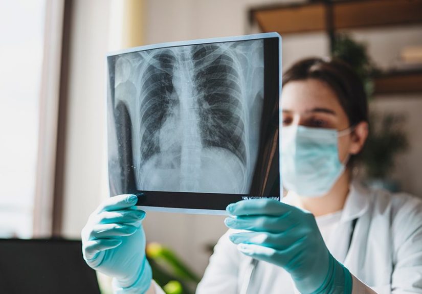
Imaging Tests
Imaging helps doctors understand the size, location, structure, and behavior of a chest wall mass. An X-ray may show bone changes, rib destruction, or calcification. Computed tomography, better known as CT, provides detailed cross-sectional images and is especially useful for evaluating bone involvement and the relationship between the tumor and nearby organs.
Magnetic resonance imaging, or MRI, is often helpful for soft tissue detail. It can show whether a tumor involves muscle, nerves, blood vessels, cartilage, or the inner chest cavity. PET/CT may be used when doctors need to evaluate metabolic activity or look for disease elsewhere in the body.
Biopsy
Imaging can strongly suggest what a tumor might be, but a biopsy is often needed to confirm the diagnosis. During a biopsy, a sample of tissue is removed and examined by a pathologist. This step is crucial because two tumors can look similar on scans but require very different treatment plans.
Biopsy planning matters. For suspected sarcoma, the biopsy path should be carefully chosen so it does not interfere with later surgery. This is one reason patients are often referred to specialized centers before any procedure is done. A poorly planned biopsy can turn a neat surgical problem into a messy puzzle, and nobody wants their treatment plan to resemble a junk drawer.
Staging and Additional Testing
If cancer is confirmed, staging tests help determine whether it is localized or has spread. This may include CT scans of the chest, abdomen, or pelvis; PET imaging; bone scans; or other tests depending on the tumor type. Blood tests may be used to assess general health before treatment, although they usually do not diagnose chest wall tumors by themselves.
Treatment Options for Chest Wall Tumors
Treatment depends on whether the tumor is benign or malignant, where it is located, how large it is, what tissue type it comes from, whether it has spread, and how healthy the patient is overall. There is no universal “one-size-fits-all” plan. Chest wall tumor treatment is more tailored than a custom suit, and usually far less fun to shop for.
Observation
Some small, benign, asymptomatic tumors may be monitored with regular exams and imaging. Observation may be reasonable when the tumor has classic benign features, is not growing, and is not causing pain or functional problems. The goal is to avoid unnecessary surgery while still watching carefully for changes.
Surgery
Surgery is a major treatment for many chest wall tumors, especially malignant tumors that can be removed. The goal is often complete resection with clear margins, meaning the surgeon removes the tumor along with a border of healthy tissue to reduce the risk of recurrence.
Depending on the tumor’s location, surgery may involve removing part of a rib, multiple ribs, cartilage, sternum, muscle, or soft tissue. When a large section of the chest wall is removed, reconstruction may be needed to restore stability, protect internal organs, preserve breathing function, and improve appearance. Reconstruction may use mesh, titanium plates, custom prostheses, muscle flaps, or other materials.
A well-planned chest wall reconstruction is not just cosmetic. The chest wall moves every time a person breathes. If it is unstable, breathing can become difficult or painful. Reconstruction helps the chest wall do its job: protect, support, and move without causing trouble.
Radiation Therapy
Radiation therapy uses high-energy beams to damage cancer cells. It may be used before surgery to shrink certain tumors, after surgery to lower recurrence risk, or instead of surgery when a tumor cannot be safely removed. Radiation may also help relieve pain or control symptoms in advanced disease.
Radiation decisions depend on tumor type. Some tumors are more sensitive to radiation than others. Chondrosarcoma, for example, is often treated primarily with surgery, while Ewing sarcoma commonly involves chemotherapy and may include radiation as part of the treatment plan.
Chemotherapy
Chemotherapy uses medicines that travel through the bloodstream to attack cancer cells. It is especially important for tumors that are likely to spread or that respond well to systemic therapy, such as Ewing sarcoma and some high-grade soft tissue sarcomas.
Chemotherapy may be given before surgery, called neoadjuvant therapy, to shrink a tumor and treat microscopic disease. It may also be given after surgery, called adjuvant therapy, to reduce the chance of recurrence. The exact medicines depend on the tumor’s diagnosis and the patient’s health.
Targeted Therapy and Immunotherapy
For some cancers involving the chest wall, targeted therapy or immunotherapy may be considered. These treatments are more common when the tumor is a metastatic cancer from another organ, such as lung cancer, breast cancer, or melanoma. Testing the tumor for specific genetic or molecular features may help guide treatment.
Not every chest wall tumor has a targeted option, but cancer treatment is becoming more personalized. When appropriate, clinical trials may offer access to newer therapies, especially for rare sarcomas or advanced tumors.
Recovery and Follow-Up Care
Recovery depends on the treatment. After surgery, patients may need pain control, breathing exercises, wound care, physical therapy, and gradual return to activity. If ribs or the sternum are involved, healing can take time because the chest wall moves constantly. Even laughing can become an unexpected core workout, which is rude but temporary.
Follow-up care is essential. Doctors monitor for recurrence, complications, healing, breathing function, and long-term effects of treatment. Follow-up may include physical exams, CT scans, MRI, or other imaging at scheduled intervals.
Patients should contact their care team if they notice increasing pain, new swelling, fever, shortness of breath, wound drainage, or a new lump. For cancerous tumors, long-term surveillance can continue for years because some sarcomas may recur late.
When to See a Doctor
A medical appointment is recommended for any chest wall lump that grows, becomes painful, feels firm or fixed, or does not go away. People with a history of cancer should be especially cautious about new chest wall pain or swelling. Persistent localized chest pain, unexplained weight loss, night sweats, or symptoms that interfere with breathing should also be evaluated.
Seek urgent medical care for severe chest pain, trouble breathing, fainting, coughing blood, or chest pain that could be related to the heart. Not every chest symptom is a tumor, and some causes of chest pain require immediate attention.
Living With a Chest Wall Tumor Diagnosis
A chest wall tumor diagnosis can feel overwhelming because it combines scary words: chest, tumor, biopsy, surgery, oncology. That is a lot for one person to carry. The first step is getting the exact diagnosis, because “tumor” only means abnormal growth. It does not automatically mean cancer, and it does not automatically predict the outcome.
Patients can help themselves by keeping records, asking questions, and seeking care from specialists familiar with chest wall tumors. Helpful questions include: What type of tumor is it? Is it benign or malignant? Has it spread? Do I need surgery? Will reconstruction be required? Should I see a sarcoma specialist? What are the benefits and risks of radiation or chemotherapy? How often will I need follow-up scans?
Emotional support matters, too. Rare tumors can make people feel isolated. A counselor, support group, patient navigator, social worker, or trusted family member can make the process less lonely. The medical plan treats the tumor; the support plan helps treat the human being attached to it.
Practical Experiences and Real-Life Lessons About Chest Wall Tumors
Many people first notice a chest wall tumor in ordinary, almost forgettable ways. Someone may feel a small bump while showering. Another person may notice one side of the chest looks slightly different in the mirror. A runner might blame rib pain on exercise, a parent might assume soreness came from lifting groceries, and a desk worker may accuse poor posture of yet another crime. Sometimes those guesses are correct. Sometimes the symptom sticks around long enough to deserve a closer look.
One common experience is delayed evaluation because the chest wall is an easy area to injure. People bump into furniture, strain muscles, cough hard during infections, sleep in weird positions, and then wake up feeling like they wrestled a refrigerator. Because muscle and rib pain are common, a slow-growing tumor may not be suspected immediately. The practical lesson is not to panic over every ache, but to respect symptoms that persist, grow, or come with a lump.
Another real-life pattern is confusion after imaging. A scan may reveal a “lesion,” “mass,” “expansile rib abnormality,” or “soft tissue density,” which can sound terrifying before anyone explains what it means. Patients often discover that imaging is only one chapter of the story. CT may show the structure, MRI may clarify soft tissue involvement, PET may assess activity, and biopsy may provide the final name. Waiting for these steps can be stressful, but each one helps avoid guessing.
People who undergo biopsy often learn that planning matters more than they expected. For suspected sarcoma, the biopsy should be coordinated with the team that may perform surgery. This helps ensure the biopsy tract can be removed during the operation if needed. It is a small detail with big consequences, and it is one reason second opinions at experienced centers can be valuable.
Surgery experiences vary widely. A small benign tumor may require a limited operation and a relatively straightforward recovery. A larger malignant tumor may require removal of ribs, cartilage, soft tissue, and complex reconstruction. Patients often describe the early recovery as a balance between protecting the chest and learning to breathe, move, cough, and sleep comfortably again. Pain control, incentive spirometry, walking, and follow-up visits become part of the daily routine.
Breathing exercises may seem too simple to matter, but they can be surprisingly important after chest wall surgery. Because deep breaths can hurt at first, patients may breathe shallowly, which increases the risk of lung complications. Guided breathing, gentle movement, and good pain management help the lungs stay open and active. In recovery, small habits can be big medicine.
Another lesson is that appearance and body image deserve attention. Chest wall surgery may leave scars, contour changes, or reconstruction materials that feel different under the skin. Some patients adjust quickly, while others need time. Neither reaction is wrong. Asking the surgical team about expected appearance, scar care, activity limits, and reconstruction options can help set realistic expectations.
For malignant chest wall tumors, follow-up can be emotionally complicated. Scans may bring reassurance, but they can also cause “scanxiety,” the nervous waiting period before results. Many patients cope by scheduling scans early in the day, bringing someone to appointments, writing down questions in advance, and avoiding late-night internet spirals. The internet is useful, but at 2:00 a.m. it can turn one symptom into a twelve-tab disaster.
Families and caregivers also have a role. Practical help with transportation, meals, medication schedules, school or work communication, and household tasks can reduce stress. Emotional support does not require perfect words. Often, the best support is simply showing up, listening, and not trying to solve everything in one heroic speech.
The biggest experience-based takeaway is this: chest wall tumors require clarity, patience, and specialized guidance. A lump is not a diagnosis. A scan is not the whole story. A biopsy result is not the end of the conversation. With the right team, many patients can receive a thoughtful plan that addresses the tumor, preserves breathing function, supports recovery, and helps them return to daily life with more confidence.
Conclusion
Chest wall tumors are uncommon, but they cover a wide range of conditions, from harmless benign growths to aggressive cancers that require complex treatment. The most important step is an accurate diagnosis. Imaging, biopsy, and specialist evaluation help determine whether the tumor is benign or malignant, where it began, and what treatment is most appropriate.
Treatment may involve observation, surgery, chest wall reconstruction, radiation therapy, chemotherapy, targeted therapy, immunotherapy, or a combination of approaches. Because the chest wall protects vital organs and supports breathing, care is often best managed by a multidisciplinary team with experience in thoracic tumors and sarcomas.
If you notice a persistent chest wall lump, unexplained swelling, worsening localized pain, or symptoms that do not make sense, do not ignore them. Most chest symptoms are not cancer, but getting checked is the only way to know. Your chest wall has an important job. When it raises a red flag, it deserves a proper conversation with a medical professional.
Note: This article is for general educational purposes only and should not replace medical advice, diagnosis, or treatment from a qualified healthcare professional.