Table of Contents >> Show >> Hide
- What You’ll Learn
- What Is the Abdomen, Exactly?
- Layers of the Abdominal Wall (Outside to Inside)
- Abdomen Area Maps: Quadrants, Regions & Diagram
- What’s Inside: Major Abdominal Organs (and Typical Locations)
- The Peritoneum: The Slippery Lining That Keeps Things Moving
- Blood Supply, Nerves, and Why Pain Can Feel “Off”
- Real-Life Anatomy: Pain Patterns, Hernias, and Imaging
- Experiences Related to Abdomen Anatomy, Area & Diagram (Extra Section)
- SEO Tags
The abdomen is basically your body’s “utility room”: it stores, processes, filters, fuels, and occasionally reminds you (loudly) that you ate too fast.
Anatomy makes it less mysteriousespecially when you learn how clinicians map the belly into predictable areas, like a GPS for organs.
What Is the Abdomen, Exactly?
In everyday speech, “abdomen” usually means the front of your belly. In anatomy, it’s more specific:
the abdomen is the part of the trunk between the thorax (chest) and the pelvis.
Think of it as the middle “housing unit” for digestion, filtration, and major blood vessels.
Key boundaries (the mental picture)
- Top: The diaphragm (a dome-shaped breathing muscle) forms the roof.
- Bottom: The abdomen blends into the pelvic cavity (no hard wall between them).
- Front & sides: The abdominal wall (muscles + fascia) wraps like a supportive corset.
- Back: The spine and deep back muscles form the posterior boundary.
You’ll also see the term abdominopelvic cavity, which simply groups the abdominal and pelvic cavities together,
because your organs didn’t install a “close door” button between them.
Layers of the Abdominal Wall (Outside to Inside)
If you could peel the abdomen like an onion (please don’t), you’d pass through several consistent layers.
Understanding these layers helps explain everything from belly-button hernias to why bruises spread the way they do.
1) Skin and superficial fascia: the “packing foam”
Under the skin sits superficial fascia, which often gets described as two parts:
a more fatty layer (commonly called Camper’s fascia) and a deeper, more membranous layer (often called Scarpa’s fascia).
The names sound fancy, but the concept is simple: one layer is squishier, the other is more sheet-like.
2) Muscle layers: the core that moves and protects
The anterolateral (front/side) abdominal wall is famous for three flat muscle layers plus a “six-pack” muscle.
Their fibers run in different directions, like crisscrossed tapegreat for strength and controlled motion.
- External oblique: fibers generally run “hands-in-pockets” direction.
- Internal oblique: fibers run mostly perpendicular to external oblique.
- Transversus abdominis: fibers run mostly horizontaldeep “belt” for stability.
- Rectus abdominis: vertical muscle inside the rectus sheath; helps flex the trunk and compress the abdomen.
3) The rectus sheath and linea alba: nature’s zipper
The flat muscles turn into broad tendons (aponeuroses) that wrap around the rectus abdominis, forming the rectus sheath.
In the midline, these tendons meet at the linea albaa strong fibrous seam that’s basically your body’s built-in centerline.
4) Deep fascia, fat, and the “inner lining”
Deeper layers include connective tissue and varying fat. Closest to the organs is the parietal peritoneum
(the body’s smooth inner lining of the abdominal cavity). Between the wall and organs you’ll also find potential spaces where fluid, air,
or infection can spreadone reason anatomy diagrams obsess over “compartments.”
Memory trick: The abdominal wall is less like one thick slab and more like a layered jacket: outer cover, insulation, cross-stitched support, and an inner lining.
Abdomen Area Maps: Quadrants, Regions & Diagram
Doctors and anatomy books divide the abdomen into standardized sections to describe findings quickly.
Instead of “it hurts somewhere near my left-ish middle-ish area,” you get a repeatable map.
The 4-quadrant system (fast and clinically common)
The abdomen is split by a vertical line down the middle and a horizontal line through the belly button:
RUQ, LUQ, RLQ, LLQ.
The 9-region system (more detailed)
This system uses two vertical lines (roughly mid-clavicular) and two horizontal lines to create nine boxes.
It’s great for pinpointing structures and describing tenderness, masses, or imaging findings.
Both maps are “correct.” The 4-quadrant map is faster; the 9-region map is more precise.
Anatomy is the rare subject where having two different maps is genuinely helpful and not just an excuse to sell more textbooks.
What’s Inside: Major Abdominal Organs (and Typical Locations)
Organ positions vary slightly by body shape, posture, and how full your stomach and intestines are.
Still, there are dependable patternsuseful for basic understanding, clinical exams, and reading diagrams.
Quadrants: “Who lives where?” (high-level)
-
RUQ: much of the liver, gallbladder, part of the duodenum,
head of the pancreas, and the right kidney (more posterior). -
LUQ: stomach, spleen, part of the liver (left lobe),
body/tail of the pancreas, and the left kidney (posterior). - RLQ: cecum and appendix, lower small intestine, and nearby pelvic structures.
- LLQ: sigmoid colon, descending colon region, lower small intestine, and nearby pelvic structures.
Regions: more detail without getting lost
The nine regions help you describe “central upper” versus “side middle” areas.
For example:
- Epigastric: stomach area, part of liver, pancreas region.
- Umbilical: small intestine loops; portions of colon.
- Hypogastric (pubic): bladder region and pelvic organ neighborhood.
- Right/left hypochondriac: liver (right), spleen (left), plus stomach/colon edges.
- Right/left lumbar: ascending/descending colon regions; kidneys are deeper and more posterior.
- Right/left iliac (inguinal): cecum/appendix region (right) and sigmoid colon region (left).
Intraperitoneal vs. retroperitoneal: why some organs “feel” different
Some abdominal organs sit inside the peritoneal sac (intraperitoneal) and are relatively mobile.
Others sit behind the peritoneum (retroperitoneal) and are more “fixed” against the back wall.
This matters for how disease spreads, how pain is perceived, and how surgeons and radiologists describe anatomy.
- Typically intraperitoneal: stomach, liver, spleen, much of small intestine, transverse and sigmoid colon.
- Typically retroperitoneal: kidneys, adrenal glands, aorta/IVC, much of duodenum, parts of pancreas, ascending/descending colon.
The Peritoneum: The Slippery Lining That Keeps Things Moving
The peritoneum is a thin, smooth membrane that lines the abdominal cavity and covers many abdominal organs.
It’s designed for low frictionso your organs can glide as you breathe, move, and digest.
Parietal vs. visceral peritoneum
- Parietal peritoneum: lines the inner abdominal wall.
- Visceral peritoneum: wraps around organs (where applicable).
Omentum and mesentery: anatomy’s “suspension system”
If the peritoneum were a fitted sheet, the mesentery would be the folds that anchor and support the intestines,
carrying blood vessels, nerves, and lymphatics to the gut.
The greater omentum hangs from the stomach like an apron over the intestines (often described as protective),
while the lesser omentum connects the stomach/duodenum region to the liver.
Diagram-reading tip: When a diagram shows a “fold,” assume it’s not just decorationfolds usually mean vessels and nerves are traveling inside.
Blood Supply, Nerves, and Why Pain Can Feel “Off”
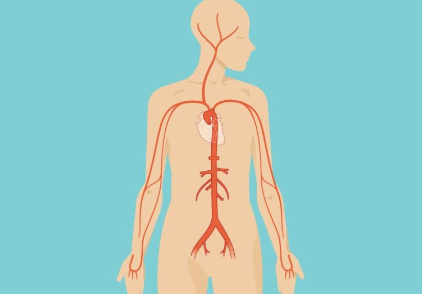
Major blood highways: aorta, celiac trunk, and mesenteric arteries
The abdominal organs are fed by branches of the abdominal aorta. Three unpaired branches are the celebrity trio:
the celiac trunk (upper abdominal organs like stomach, liver, spleen),
the superior mesenteric artery (much of small intestine and proximal colon),
and the inferior mesenteric artery (distal colon).
Venous blood returns via large veins including the inferior vena cava, while much of the digestive tract drains through the
portal venous system to the liver for processing.
Somatic vs. visceral pain: “point with a finger” vs. “wave at the whole area”
The abdominal wall and parietal peritoneum have somatic (body wall) nerve supplypain tends to be sharper and more localized.
Many internal organs produce visceral pain that can be dull, crampy, and harder to pinpoint.
That’s also where referred pain can show upyour brain interprets signals in familiar “maps,” sometimes projecting discomfort
to a nearby region rather than the exact structure.
Why diagrams matter here
When clinicians document “RUQ tenderness” or “epigastric discomfort,” they’re using the area maps to communicate quickly.
It doesn’t magically diagnose a cause by itself, but it narrows the neighborhood of possibilities and guides the next steps
(exam maneuvers, labs, and imaging choices).
Real-Life Anatomy: Pain Patterns, Hernias, and Imaging
Anatomy becomes instantly practical when you connect structure to common situations.
Here are a few anatomy-grounded examples (educational onlynot a diagnosis guide):
1) Abdominal wall vs. “inside” pain
Pain from the abdominal wall (muscle strain, irritated nerves, scar sensitivity) often feels more superficial and may worsen with
movements that tighten the corelike sitting up, coughing, or twisting.
Deeper organ-related discomfort can feel more diffuse and may come with general symptoms like nausea or changes in appetite.
2) Why hernias happen where they do
A hernia occurs when tissue pushes through a weak spot in the abdominal wall.
The belly button (umbilicus), previous surgical incisions, and groin regions are common “stress points” because the wall’s layers
naturally change thereor were interrupted and healed.
3) Imaging “chooses the neighborhood first”
In many clinical settings, the location of symptoms helps determine which imaging is most useful.
Ultrasound is often used for certain upper-right abdominal questions, while CT is commonly used for broader or lower abdominal concerns.
The reason is practical: different tests visualize different structures better, and anatomy guides that decision.
4) Surface landmarks you’ll see in diagrams
- Costal margin: the lower edge of the rib cageoften used to describe upper abdominal locations.
- Umbilicus: the belly buttoncommonly used as a reference point for the quadrant cross.
- Iliac crests: the “top of the hips”useful when describing lower abdominal regions.
- Midline (linea alba): the central seamimportant for anatomy diagrams and surgical approaches.
If you remember one thing: abdominal diagrams aren’t trying to be artthey’re trying to be consistent. Consistency is what turns “belly” into a usable medical map.
Experiences Related to Abdomen Anatomy, Area & Diagram (Extra Section)
Even if you never take a formal anatomy class, you’ll still “experience” abdominal anatomy in daily lifethrough movement, digestion, posture,
and the way people describe sensations. This is one of those topics where diagrams stop being abstract the moment you connect them to real moments.
A common experience: someone says, “It hurts right here,” and they point to a spot with one or two fingers.
That simple gesture is basically an anatomy diagram in human form. People naturally draw a map on themselves: upper vs. lower, right vs. left,
central vs. side. Clinicians do the same thingjust with standardized words like “right upper quadrant” or “epigastric region”
so everyone on a care team imagines the same location. When you learn the quadrant and region diagrams, you start noticing how often daily language
already tries to do this (we just do it with less precision and more dramatic hand-waving).
If you’ve ever done core exercisesplanks, sit-ups, dead bugs, even laughing too hard at the wrong timeyou’ve met the abdominal wall muscles.
The “burn” you feel isn’t random; it’s the rectus abdominis and the layered obliques and transversus abdominis doing their jobs:
stabilizing your trunk, managing pressure, and coordinating with breathing.
Many people are surprised to learn that the deepest layer (transversus abdominis) is less about showing off and more about supportlike a wide belt
that firms up your midsection when you lift, twist, cough, or even sing loudly in the car.
Once you know the layers, fitness cues like “brace your core” suddenly sound less like motivational poetry and more like anatomy.
Digestion is another everyday anatomy lesson. After a big meal, you may feel fullness high in the abdomen, more central, sometimes described as
“upper belly” discomfort. That matches the region diagrams: the stomach sits largely in the upper central/left area, while loops of small intestine
occupy much of the middle abdomen, and the large intestine frames the outer edges like a picture frame.
It’s also why sensations can shift as digestion progressesgas and movement travel, and the “where” you feel it can migrate.
Understanding the basic organ layout doesn’t replace medical evaluation, but it does help you interpret what your body is trying to communicate.
Students who learn abdominal anatomy often remember it best through stories and pictures: the “apron” of the omentum, the “zipper seam” of the linea alba,
the layered directions of the obliques like crossed tape. Many also remember the abdomen as a set of neighborhoods:
RUQ is “liver/gallbladder territory,” LUQ is “stomach/spleen territory,” and the lower quadrants are “intestine and pelvic neighborhood.”
Once those neighborhoods stick, diagrams become easier to read. Instead of memorizing a list, students start asking better questions:
“Is this organ intraperitoneal or retroperitoneal?” “What vessel supplies this area?” “If pain is vague and central, is it more likely visceral?”
Those are anatomy-fluent questionsand they’re exactly what makes diagrams useful rather than decorative.
One more very human experience: posture and breathing. The diaphragm (top boundary of the abdomen) moves with every breath, and your abdominal wall
respondssubtly tightening and relaxing. When you take a deep breath, your abdomen can expand because pressure shifts and the diaphragm descends.
When you cough or laugh, abdominal muscles contract to help manage pressure. That’s anatomy you can feel in real time.
And once you’ve seen an abdomen diagram, it’s hard not to imagine those moving parts working together like a coordinated team:
diaphragm above, pelvic floor below, abdominal wall wrapping around, and organs gliding on smooth linings in between.
In other words: the abdomen is not just “where food goes.” It’s a dynamic, moving systemmore like a well-organized workshop than a simple storage bin.